Lab study: Japanese chemists use cancer cells' own carbon monoxide to make a drug inside them
Researchers at RIKEN and the Institute of Science Tokyo have designed a palladium compound, carried inside the blood protein albumin, that reacts with the extra carbon monoxide made by cancer cells to produce a toxic anticancer molecule on the spot. The method has so far been tested only on cells grown in the lab, and the authors describe its cell-killing effect as modest. It is a proof of concept, not a treatment.
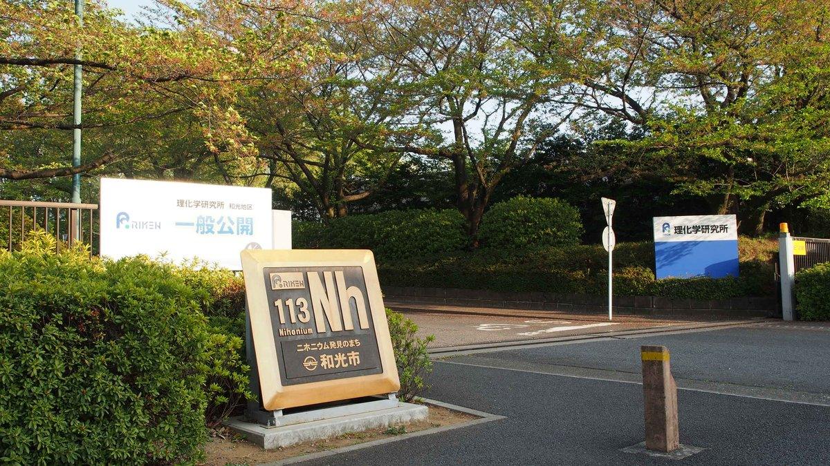
Many cancer cells make more carbon monoxide (CO) than healthy cells, because an enzyme called heme oxygenase-1 is more active in them. A team led by Katsunori Tanaka at the RIKEN Pioneering Research Institute in Wako, near Tokyo, and the Institute of Science Tokyo has tried to turn that by-product against the tumour: instead of delivering a finished drug, they deliver a harmless precursor that only becomes toxic when it meets the extra CO inside a cancer cell. The study, by Masayuki Kawai, Tsung-Che Chang, Ambara R. Pradipta and Tanaka, was published on 17 September in the open-access journal Nature Communications. RIKEN presented the results on 23 October.
The chemistry relies on palladacycles, palladium compounds that can capture CO and turn it into phenanthridinone, a molecular core found in some drugs that block the enzymes PARP and topoisomerase. The problem is that inside a cell, the palladium is quickly disabled by glutathione and similar sulphur-containing molecules. The researchers solved this by tucking the palladium compound into a pocket of human serum albumin, a common blood protein, using a coumarin "anchor" to hold it in place. The albumin shields the metal and also helps the package get into cells.
In a first test, the team used a fluorescent version of the system on four human cancer cell lines, from liver (HepG2), cervix (HeLa S3), breast (MCF7) and lung (A549) cancers, and on normal TIG3 fibroblasts. After 48 hours, the product had formed in all the cancer cells, from 59 to 136 picomoles per million cells (77 in the liver cancer cells), against only 15 in the normal cells.
The team then built a version that releases a known type of anticancer molecule, a phenanthridinone related to the natural compound oxynitidine that inhibits the enzyme topoisomerase I. In HepG2 liver cancer cells, the drug was detected inside the cells and the treatment slowed their growth. It was about three times more toxic to the liver cancer cells than to the normal fibroblasts, and blocking the cells' CO-producing enzyme weakened the effect, which supports the idea that the cells' own CO is the trigger. In a 12-day test, it also reduced the growth of breast cancer cell colonies.
A possible risk is that the drug could be made too early, in the blood, which also contains CO. In a test in fresh mouse blood kept at 37°C for four hours, the researchers detected no product, probably because the CO in blood is tightly bound to haemoglobin. This was a test-tube check of blood samples, not an experiment in living animals.
The authors are cautious about the results. They write that the toxic effect is "modest" and that the difference between cancer and normal cells is a "modest therapeutic window", and they describe the work as a proof of concept. All the cancer experiments were done in cell cultures; the method has not been tested as a treatment in animals or people. According to RIKEN, the next step is to move from cell lines to animal models. "This work represents an important advance in therapeutic in vivo synthetic chemistry," Tanaka said in the institute's statement.